A cleft is a congenital deformity of the lip, palate, or face. Multiple surgeries have to be performed from birth for a cleft baby/patient to get into a normal life aesthetically and functionally. So here is one of our patients, with his journey of transformation from Cleft Maxillary Hypoplasia / severe maxillary retrognathism to normal facial appearance.
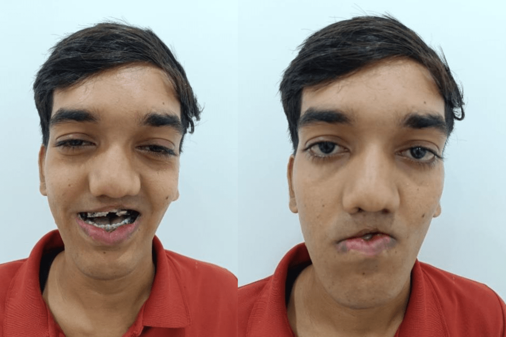
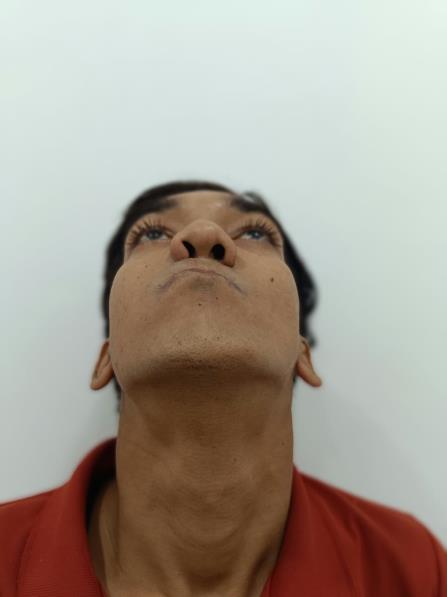
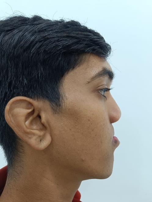
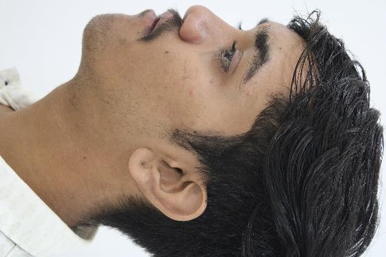
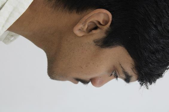
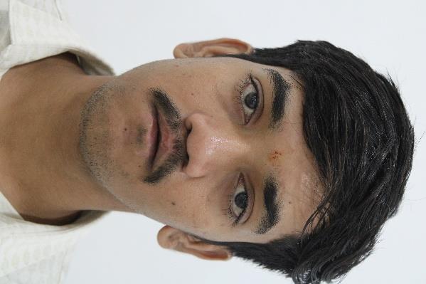
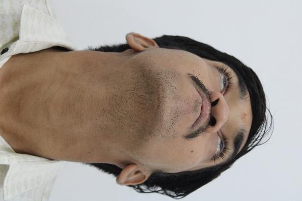
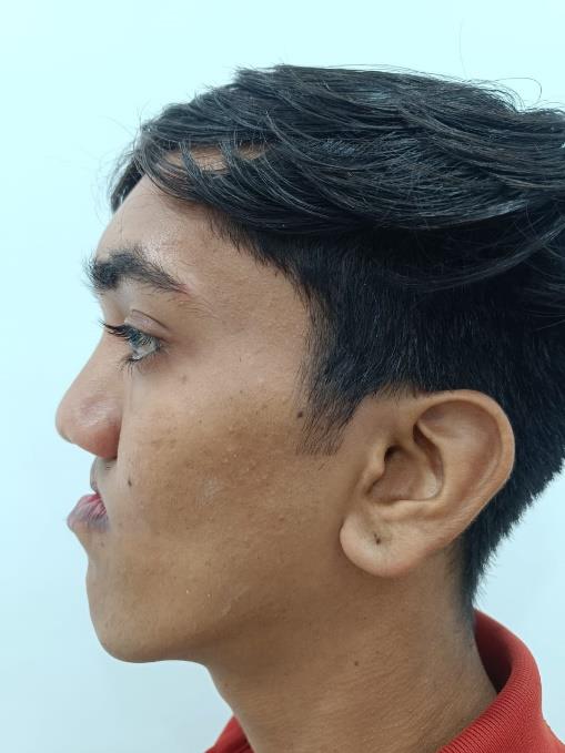
The following images show front, side, and worm’s view of the patient before surgery.
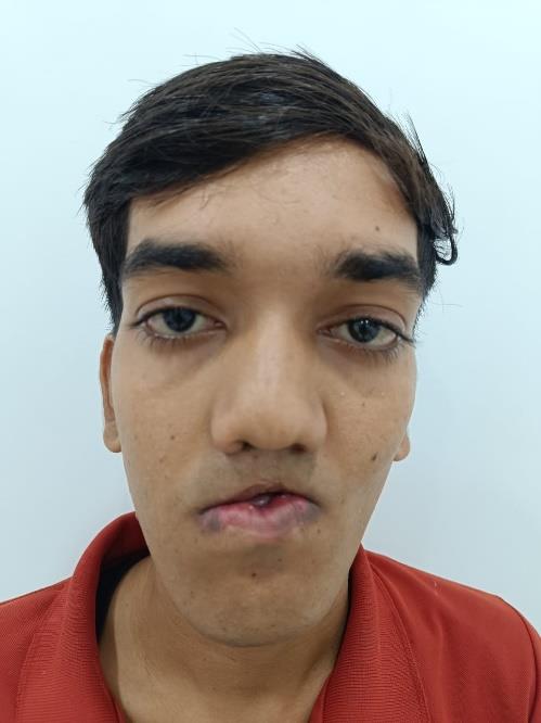
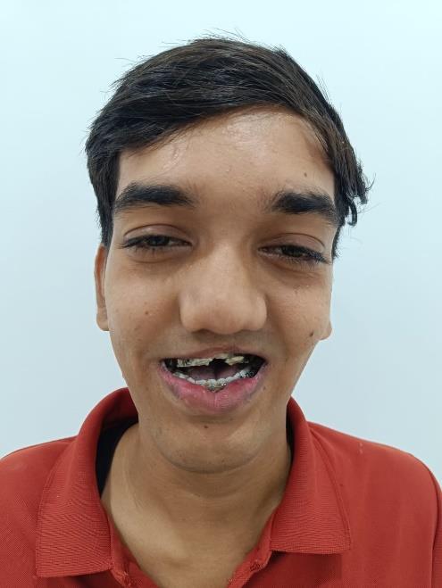
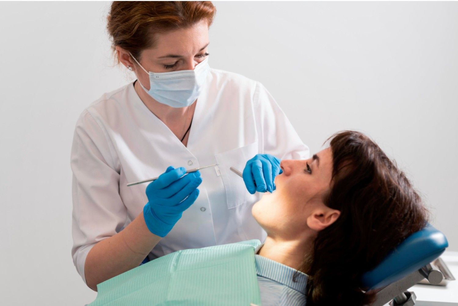
This patient had consulted many other hospitals to correct his deformity but couldn’t achieve the desired results and was finally referred to us. We performed a detailed facial and skeletal analysis using CBCT (Cone Beam Computed Tomography), intra-oral and extra-oral photographs, dental scans, and analysis. Severe Cleft Maxillary Hypoplasia with maxillary retrognathism/underbite was noted, and we decided to proceed with an Anterior Maxillary Distraction procedure.
Anterior Maxillary Distraction Procedure
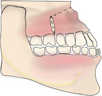
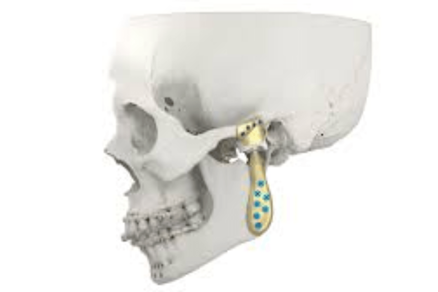
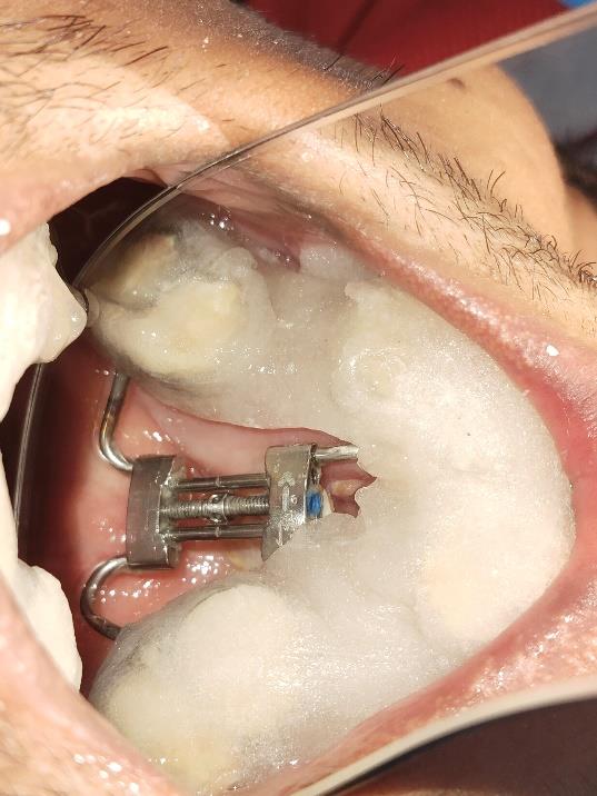
It is a surgical procedure to correct deep underbite and maxillary deficiency in Cleft Maxillary Hypoplasia patients, where a tooth-borne device called the AMD device is fixed inside the mouth prior to surgery.
AMD TOOTH BORN DEVICE
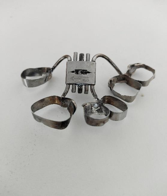
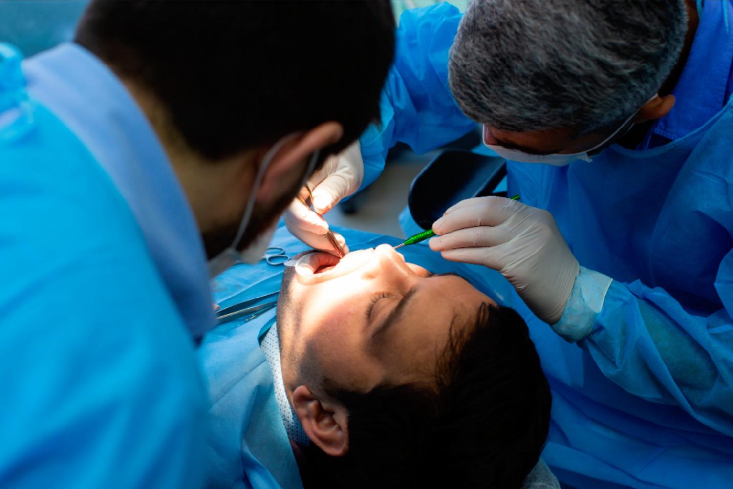
In surgery, the anterior maxilla is mobilized using surgical cuts on both sides between the premolars and molars, and the tooth-borne device is activated 2 to 3 days after surgery by daily turning, so the anterior maxilla slowly progresses in a forward direction.
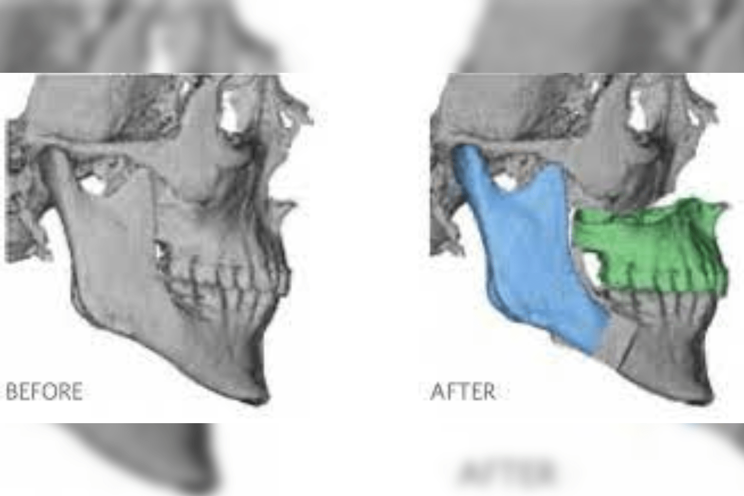
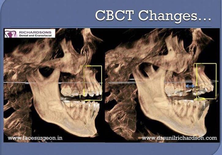
The following image shows the CBCT changes happening during the activation of the AMD device post-surgery. In patients with Cleft Maxillary Hypoplasia, up to 13 mm of advancement can be achieved depending upon the patient’s requirement, making AMD an effective treatment option for Cleft Maxillary Hypoplasia cases.
Once the advancement is achieved to the planned direction, to stabilize the results A BUNK has to be placed iunside the mouth.
After that, the patient can go back to his normal life with some dietary restrictions. Follow-up of the patient was done 3 months post-surgery.
Again, CBCT was taken to assess the bone formation on the surgical site and the advanced area of the maxilla. In cases of Cleft Maxillary Hypoplasia, if the bone formation is good enough, the bunk can be removed along with the device.
We recalled him after 4 to 5 months and assessed his bone formation by taking CBCT. The healing was excellent, and we removed the bunk and the tooth-borne device. Complete intraoral cleaning and polishing was done, marking a successful correction of Cleft Maxillary Hypoplasia.
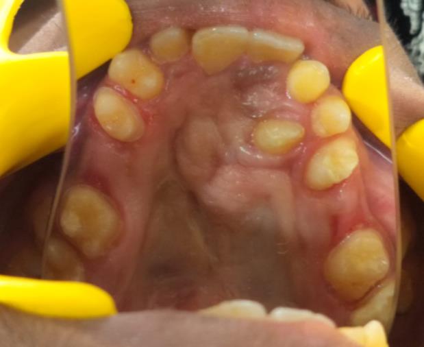
IMMEDIATELY AFTER THE BUNK REMOVAL
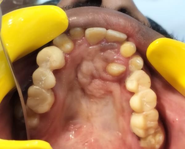
This was the results we achieved immediately after the bunk removal.We can clearly appreciate the advancement that we have achieved through this procedure,the archform is satisfactory,the underbite is
corrected.
In this stage we will be giving either an acrylic plate or temporary resin bridge bilaterally to retain or stabilize the result so it will ensure the further growth and calcification of the bone
Temporary bridge bilaterally to stabilize the result
A remarkable improvement in facial appearance can be seen in this stage itself,and our patient was very much thrilled to see his result,as it was improved his functionality and also his aesthetic considerations.
EXTRAORAL IMAGES SHOWING THE RESULTS.
After this stage ,dental rehabilitation such as SMILE DESIGNING has to be planned to enhance his facial appearance also to improve his functionality.
This is the beautiful journey of our beloved patient, who has come to us with lots of expectations and a very humble and co-operative patient of us ,and we are glad to share his journey and transformation from a cleft hypoplasia to almost a normal facial appearance.He was very excited about his new face, and we are glad that we are part of his happiness.

The before and after treatment images of the patient showing, the advancement of maxilla with the surgery.