Introduction
Vascular lesions, more commonly known as Haemangioma, are anomalies involving an abnormal growth of blood vessels. These hemangiomas of the face can range from benign conditions, such as Hemangiomas and vascular malformations, to more complex and potentially life-threatening lesions, like Arteriovenous Malformations (AVMs).
Classification
Vascular lesions, including hemangiomas of the face, are classified into vascular malformations and vascular tumours:
Vascular malformations –
These are lesions arising from abnormal development of blood vessels during embryogenesis. Unlike tumours, they do not exhibit rapid growth but instead grow proportionally with the individual. Different types of vascular malformations include arteriovenous malformations, capillary malformations, venous malformations, and lymphatic malformations.
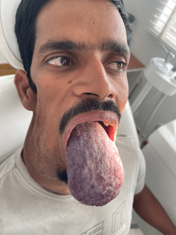
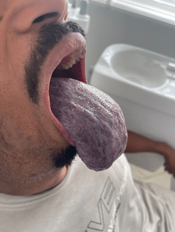
Arteriovenous malformations are high-flow with a characteristic sound called bruit and are pulsatile in nature. Venous malformations are low-flow, bluish masses and often contain calcified blood clots called phleboliths. Lymphatic malformations, like hemangiomas of the face, may also present as soft, compressible lesions that can cause swelling and deformity in affected regions.


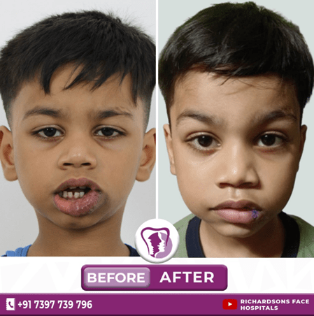
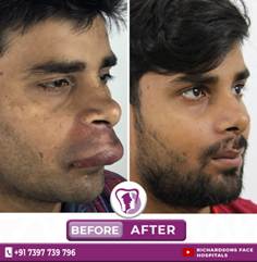
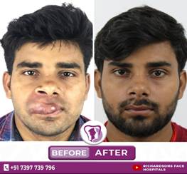
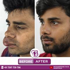
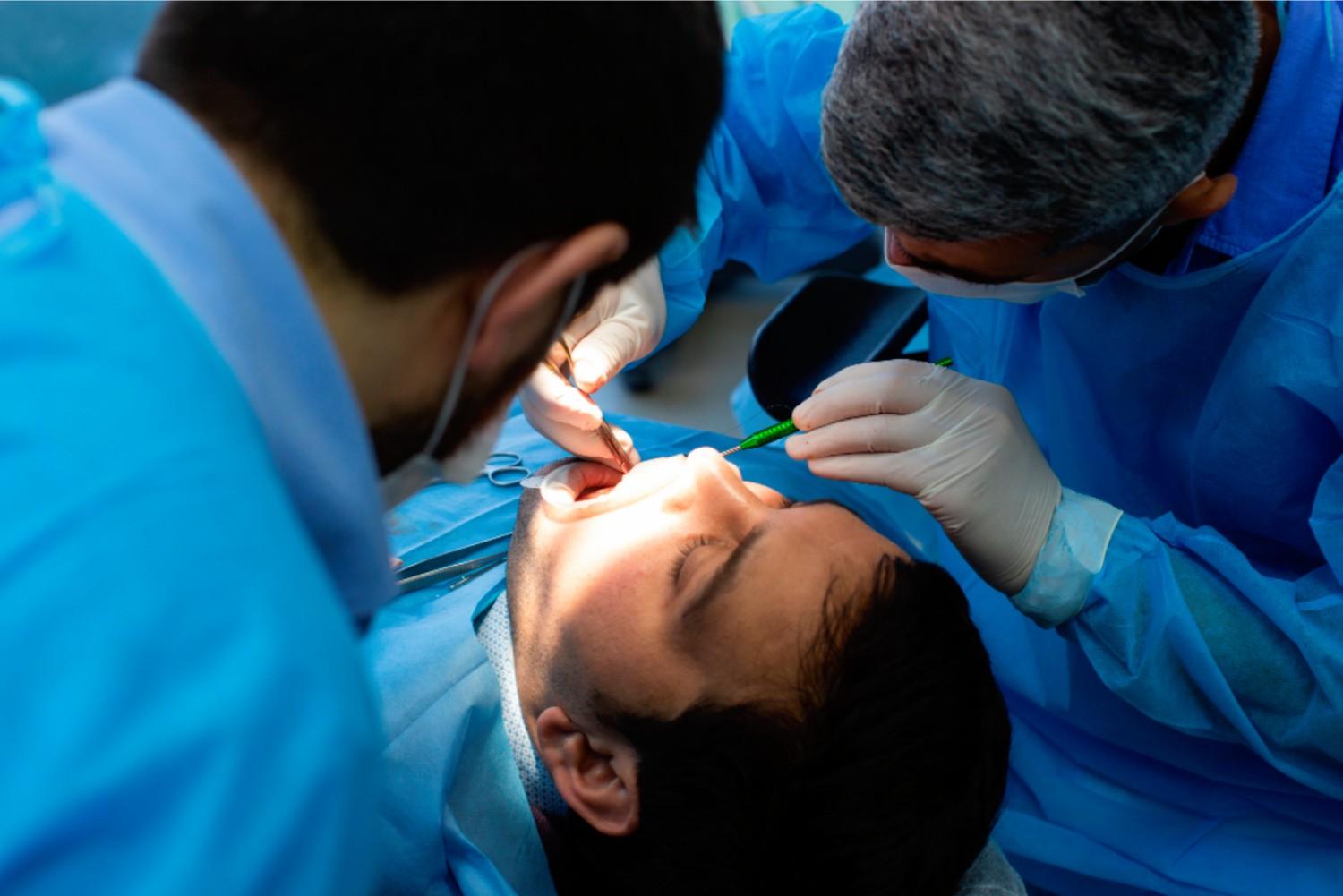
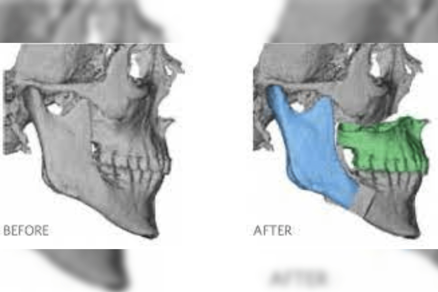
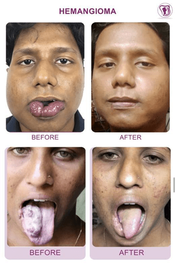
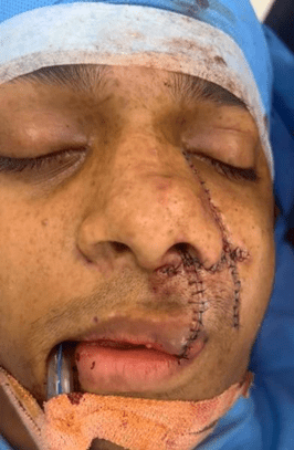
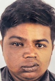
Involuted haemangioma lesions of the face before and after surgical excision
Clinical features of vascular lesions
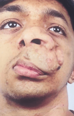
- Cosmetic deformities-
Port-wine stains or haemangiomas can cause significant cosmetic concerns.
Port wine stain
Patients with hemangiomas of the face or other visible vascular lesions may experience psychological distress, social stigma, and low self-esteem.
In many cases, haemangiomas are asymptomatic. However, in critical areas, they can cause vision problems if located near the eyes, airway obstruction if on the neck near the respiratory tract, and functional impairment due to facial distortion or restricted movement.
AV malformations may cause pain if located in deeper tissues, while vascular malformations may cause swelling that presents as soft, compressible, or spongy masses.
Hemangiomas of the face can also lead to bleeding — high-flow lesions like AV malformations are prone to spontaneous or trauma-related bleeding, while low-flow malformations such as venous malformations may bleed if severely injured or ulcerated.
Diagnosis of vascular lesions
- Clinical examination with a detailed history is essential for identifying the type, location, and extent of the lesion
- Imaging studies, such as ultrasound,d helps evaluate the depth and blood flow
- MRI or MRA provides detailed information about these signs and extent of the lesion and the blood supply to the lesion
- Biopsy is done in cases where the diagnosis is unclear
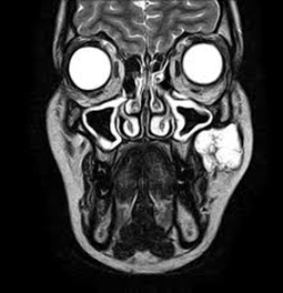
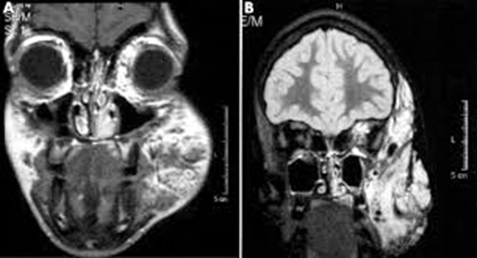
MRI of the face showing vascular malformation
Treatment of vascular lesions
Observation-
Many vascular malformations, particularly small and asymptomatic ones, simply require monitoring over time. Hemangiomas of the face, particularly in infants, often undergo involution without treatment
Medical treatment-
- Propranolol and corticosteroid therapy is used to reduce the size and vascularity of the hemangioma by inhibiting new blood vessel formation and promoting involution. They also promote fibrosis of the lesion.
- Sclerotherapy involves multiple injections at regular intervals or until the lesion regresses in size either prior to or post surgical excision.
- Sometimes it is the only treatment modality followed for lesions and good results are seen
Surgical treatment-
- Debulking of the lesion refers to removal of part of the lesion, usually done when the lesion is very large or is close to a critical structure or when complete excision of the lesion is not possible in one surgery.
- Surgical excision of the lesion refers to complete removal of small to moderately sized lesions
- In some hemangiomas of the face, excision may be considered for residual lesions that persist after involution.
Embolization-
- For high-flow lesions like AVMs, embolization involves introducing substances into the blood vessels to block/reduce the blood flow to the abnormal vascular channels.
- This then reduces the size and vascularity of the malformation.
- This procedure is usually done by an interventional radiologist and most often in conjunction with surgical excision of large tumours.
Laser therapy-
- Pulsed dye lasers are particularly effective for superficial capillary malformations such as port wine stain and help reduce redness and the appearance of the lesion
Complications
- Bleeding, particularly in high-flow lesions
- Infection
- Functional impairment when lichens are located near vital structures it can lead to breathing difficulties, visual disturbances or swallowing problems
- Cosmetic deformities, visible lesions
Prognosis
Most vascular lesions, if managed appropriately, have a good prognosis, but cosmetic outcomes vary according to the lesion.